Cosmic Baby , Magnetic Protein —Cosmic connection to Human : A possibility
Ramen Kumar Parui * 
1ARC, Room No – F101, Block – F, Mall Enclave, K. B. Sarani, Kolkata, West Bengal India .
http://dx.doi.org/10.13005/OJPS10.01.11
Protein crystal plays an important role in creation of human eyes i.e. high quality protein crystallization ensures the formation of beautiful eyes in human faces as well as ageless appearance in body structure. Cosmic Baby , i.e. the Swift J1818.0 – 1607, provides a possible clues to the scientists in the form of “Triaxiality” for creation of beautiful eyes. This author suggests that the “triaxiality” phase appears at the early stage of birth when human embryo (inside the mother’s womb) faces the effect of change in shape of the gestational sac (that covers the human embryo ) from circular shape to ellipsoidal shape. Effect of magnetic field and its magnetization force offer an opportunity to the scientists for understanding the physics of creation of high quality protein crystals. To ensure the exact role of “Triaxiality” towards beautiful eye formation needs a lot of investigations on crystallization of protein in strong magnetic field as well as finding the link of cosmic connection with human beautiful eyes.
Copy the following to cite this article:
Parui R. K. Cosmic Baby , Magnetic Protein—Cosmic connection to Human: A possibility. Oriental Jornal of Physical Sciences 2025; 10(1).
DOI:http://dx.doi.org/10.13005/OJPS10.01.11Copy the following to cite this URL:
Parui R. K. Cosmic Baby , Magnetic Protein—Cosmic connection to Human: A possibility. Oriental Jornal of Physical Sciences 2025; 10(1).Available here: https://bit.ly/42lQWfF
Download article (pdf) Citation Manager Publish History


Introduction
The main function of proteins in living organisms including human are : (a) controlling of almost all the biological reactions, and (b) to maintain a deep relationship through their structure and function which are related to the development of life 1 . In one side it is considered that neighboring atom formation 2 are the main structural basis in many biological systems while in other side the attached hydrogen atoms take an active part in playing biological functions, such as mechanism of enzyme production 3,4 . In deep sense, the precise location of this hydrogen atoms is very much crucial in order to understand the physics of origin or mechanism related to the concerned biological problems by maintaining a relationship with solid state physics5,6 .
The unknown crystal structure , in general, is resolved through the conventional method , i.e. the X-ray diffraction technique 7,8 but the problem arises due to the interaction of X-rays with the electrons of the atoms located inside the crystal structure. An alternate is to consider heavy atoms having neutrons with its isotropic form or other so that the determination of the crystal structure is possible based on the relative position of a hydrogen atom located with reference to the known presence of relatively heavy atoms 9,10. Therefore, control of physical parameters are essential for obtaining accurate result. Various methods have been developed in this regard by controlling the physical parameters such as the temperature 11, hydrodynamic field 12, electric field 13, magnetic field 14 and electromagnetic field 15. Out of these, magnetic field method has a great potential for understanding the protein crystal structure and its growth.
Although it is uncertain yet scientists believe that the knowledge of protein were known during the period in the 19th and early 20th Century. In 1938 Berzelius first cointed the term “Protein” but remained unpopular under the influence of more popular terms like “proteid”, “albumineous”, “colloids” on that time. Looking back into the past the available literature indicates that in 1840 Friedrich Ludwig Hünefeld first observed protein crystal serendipitously in a sample of dried menstrual blood sticked between glass plates used for experiments in his laboratory (figure 1B ) 16 .
In fact, the word “Protein” is derived from the Greek word ‘Proteios’ which means primary / primary importance because proteins are the most important chemical substances which are essential for growth, repair and development of life. These proteins have been found in all living cells implying that they occur in every part of the body and form the fundamental basis of structure and functions of life. For example, in human body skin, hair, haemoglobin, nails, enzymes and cells, etc. all are made of proteins.
However, through gradual studies protein crystallization area developed a lot and it has become an input part of our life, in particular through which we visualize the objects in the world.
Materials and Methods
The Birth of Biocrystallogenesis Initially, protein crystal was confined its purification and then turned as instrumental tool to demonstrate its catalytic activities in the form of “enzyme” that resides with the protein itself. In the 19th Century the most of the crystalline proteins (i.e. haemoglobins and albumins) which were considered as pure substance. Because of their availability in the micro-scale. But in 1934 a breakthrough occurred in this field when John D. Bernal and Dorothy Crowfort Hodgkin (see the review Parui 2024 19 ) described the secret structure / pattern inside the protein first time through X-ray diffraction. This opened a new era in chemistry and physiology as biocrystallogenesis. Below a few landmarks are mentioned in protein crystallization.
Year | Macromolecular Form | Crystallized Entity | Source |
1840 | Globins | Haemoglobin | Human blood |
1855 | Phytoglobulins | Excelsin | Brazil Nut |
1890 | Enzymes | HEW Lysozyme | Pig |
1926 | Hormones | Insuline | Rabbit |
1935 | Viruses | Plant virus | TMV |
1937 | Enzymes | Cotalase | Beef liver |
1969 | Antibodies | Intact 1g G | Human |
1971 | Toxins | Erabutoxin | Sea Snake |
1975 | Sweet testing Proteins | Thaumalin | Thaumato cocus Daniellii |
1980 | Membrane Protein | Porin | Escheriochia Coli |
1980 | Ribosomes | - | Bacillus Stearo thermophiles |
1984 | Protein DNA Complexes | Nucleosome | Xenopus laevis |
1988 | DNA fragments | Synthetic DNA duplexes |
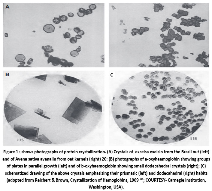 | Figure 1 : shows photographs of protein crystallization. (A) Crystals of excelsa exelsin from the Brazil nut (left) and of Avena sativa avenalin from oat kernels (right) 20: |
Protein Crystallization in Living Cells
Protein crystallization in living cell was observed surprisingly as an evidence of phenomenon for recombinant protein. It means that this micrometer-sized can be used for structural biology. For example, this in cellulo crystallization could offer us a new possibilities that do not crystallize under normal conventional approach. In other words, this unknown mode of regulation of native in cellulo crystal formation offers a challenge to the scientists. Although the mechanism of how intracellular protein self-assembled is not known yet scientists believed that the hidden regulatory mechanism ( different from normal regulatory approach ) may have a link to a specific function which is either disease associated or new form of recombinant as a protein crystallization chamber that systematically exploit living cells.
Protein Storage
In nature, nutrient storage is observed as a normal process in living objects in the form of eggs or seeds in the form of crystallize yolk proteins as a source of constant nutrient supply for the offspring. In the case of human eyes it was thought that the only 10 or so crystalline proteins ( like alpha (?), beta (?) and gamma (?) ) are involved in the evolution of eyes. The remaining lens proteins are non-conserved, diverse group used as enzymes elsewhere in the body.
Sturcture of Eye Lense
Eyes collect light through an aperture and focus it with a lens photoreceptor cells convert photons into neural signals. Lenses are of tightly packed proteins. n the case of human lenses (i.e. vertebrates) these are actually formed from modified epithelial cells containing highly concentrated soluble proteins, so called “crystallins” because of their packing arrangements into an arrays ). As mentioned above that the 10 or so crystalline proteins found in human eyes which are unique to lens tissue but present thought is of the large number of crystallins i.e. alpha (?), beta (?) and gamma (?) crystallins that are indeed also specialized lens proteins. In fact, crystallins or transparent proteins are arranged in approximately 20,000 thin concentric layers (i) whose average concentration is about twice than that of other intracellular proteins, and (2) that may play a structural role during the development of eye-lens. It means that in one side the eye lens can be used as a source to study the mechanism of aggregation and deposition of other “mis-folded” proteins that cause cataract while in another side the same eye lens provides a mechanism of aggregation of exogenous proteins whose developing approaches provides a clue how to protect the cells from the accumulation of aggregated proteins. Note that out of the earlier mentioned the 10 or so crystallins proteins alpha (?) and beta (?) crystalins are specialized lens protein that are related to heat shock proteins (in the case of vertebrates) and schistosome egg antigen, respectively. In case of the remaining vertebrates the lens proteins are a non-conserved and diverse group. This implies that one gene coding for a protein have two entirely different functions which is known as “gene sharing” and it may be a prelude a “gene duplication”
As mentioned earlier that in human eye (i.e. vertebrate) the alpha (?-), beta (?-) and gamma ( ?-) crystallins are known abundant vertebrate eye lens protein because of their presence more than 90% of total proteins. Not only that, presence of enzymes in eye lens also act as structural proteins, receptors, connexins , water channel aquaporin and cytosketal proteins. Basically, alpha (?-) crystallin,as a small heat shock protein, interacts with partly folded proteins in a chaperone manner as wll as prevents their irreversible aggregation and precipitation, beta (?-) and gamma ( ?-) crystallins are primarily composed of antiparallel beta (?-) sheet. Finally, these three i.e. the ?-, ?-, and ?-crystallins representing in all vertebrate lineages may have evolved from proteins that support this kind of filament system as well as may shed light on the functions of these proteins in lens and non-lens cells.
Eye lens Chaperon a-crystallins and Triaxial shape
The a-crystallins are molecular chaperons which belongs to the small heat shock protein family. The cellular function of this ?-crystallins is to bind to partially unfolded polypeptides and nurture them so that they are within a refolding competent state 21-23 such that their acting as molecular traps ultimately protect the cells from the consequences of irreversible protein aggregation.
There are two types ?-crystallin genes, namely, ?A and ?B . In a study of the structures of the recombinant ?-crystallins , in particular ?B-crystallins, by electron microscope 24 it was found human ?A- and ?B- crystallins were recombinantly expressed in “ Ecscherichia Coli”. Further analysis using selected 2565 single particle images (fig.2A) and Eigenimages ( i.e. images representing the most important differences in the data set (fig.4B) and preliminary class averages (fig.2C and D) they found human ?B-crystallin oligomer is roughly spherical with a diameter of 13.5 nm (fig. 3A). This oligomer also accommodates a large central cavity of
.jpg) | Figure 2: Observed image analysis of negatively stained ?B-crystallin |
 | Figure 3: 3D reconstruction of human recombinant ?B-oligomers as obtained by Peschek team. 24. |
openings at the positions of the 2-fold axes ( with diameter of ~ 3.5 nm ) and 3-fold axes (with 3 and 2 nm for the two unequals ). The overall structure of human recombinant ?B-crystallins is nearly circular based on the surface representation of 3D model (fig. 3B) and density cross section through 3D model (fig. 3B).
Aquaporin (AQP1) Water Transporter
In another investigation 25 for understanding the function of protein (i.e. 28kDa ) as water transporter in human eye-lens it was demonstrated that under the injected controlled oocyte with water in Xenopas laevis the protein was christened “ aquaporin” ( so called “AQP1 ) such that initially with a circular shape it deformed from its circular shape to deformed triaxial shape in the case of injected with cRNA. A noticeable change in shape they observed 30 seconds later from the time injection i.e. the AQP1 oocyte had begun swell as seen in figure 4 ( i.e. shape changes from circular to triaxial) and after 3 minutes the swelled AQP1 oocyte had further deformed ( i.e. from triaxial shape to ellipsoidal ) and finally exploded. This implies that the AQP1 oocyte changes its shape from initial circular to triaxial shape ( during the beginning of swelling) and finally exploded when triaxial shape turns into more deformed phase ( which was more unstable resulting which it exploded).
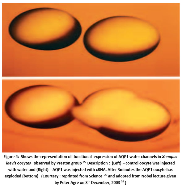 | Figure 4: Shows the representation of functional expression of AQP1 water channels in Xenopus laevis oocytes observed by Preston group 25. |
Results
Physical Observation of Hen-eggs
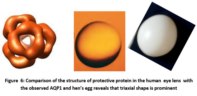
Physically observed of hen eggs by this author are shown in figure 5. A variety of egg shapes are circular (serial no 1), triaxial (sl. No 2,3,7), ellipsoidal (sl. No.4. 5, 6 ). Comparing the observed shape of 3D reconstruction of human recombinant ?B-crystallin with the physically observed shapes of hen’s egg it is revealed that the nearly circular shapes are triaxial shapes. This means that in human eye-lens proteins are associated in “Triaxial Shpes”. Of course, this needs further precise imaging of ?-crystallin to confirm it.
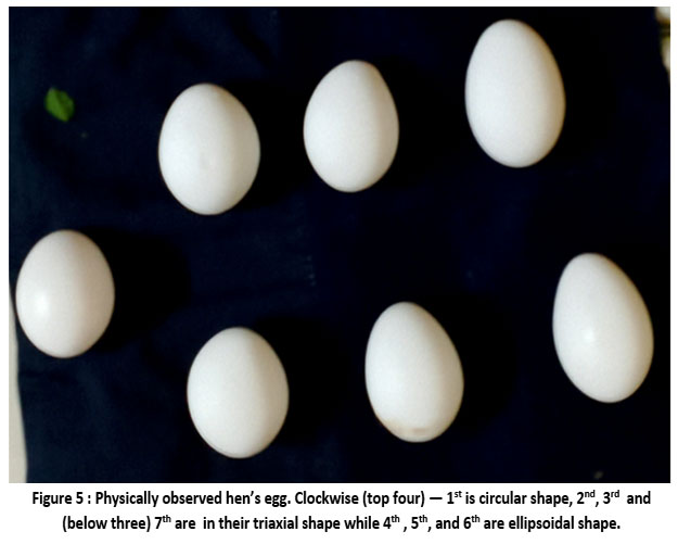 | Figure 5: Physically observed hen’s egg. Clockwise (top four) — 1st is circular shape, 2nd, 3rd and (below three) 7th are in their triaxial shape while 4th , 5th, and 6th are ellipsoidal shape. |
 |
Discovery of Cosmic Baby
During the analysis of observed X-ray data (for short burst originated from magnetar) collected by Swift X-ray telescope (XRT) Esposito team 30 discovered a typical characteristic of short burst from a magnetar on 12th March 2020 at 21:16: 49 UT 27,28. This newly discovered magnetar was actually an uncataloged X-ray source as Swift J1818.0 – 1607and presently known as “Cosmic Baby” (see fig. 7). It is called “baby” ( with age only 240 years) in the sense that till date all the discovered magnetars are about million years aged. The significant parameters of Cosmic Baby are :
Characteristic age ~ 240 years 29
Magnetic field strength at Surface ~ 2.7 x 10¹⁴ G
Magnetic field strength at poles ~ 7 x 10¹⁴ G
Spin period = 0.7333920 s
Coherent periodicity of X-ray signal = 1.36s
Period derivative ~ 9 x 10 – 11 s.s-1 30
Using the initial observed parameters at the time of discovery Parui 31- 33 estimated the internal core magnetic field inside the magnetar and it associated ellipticity ( a property measuring the deformation ) of this Cosmic Baby and found 8.9424 x 10¹⁷ G and 9 x 10 – 3 , respectively. It is seen that the estimated deformation of this object due super-strong magnetic field turns it into a “Triaxial Star”. In fact, as its age is 240 years the estimated value hints that this cosmic baby will exhibit its triaxial nature for next 760 years. The importance of cosmic baby is that it shows the possible evidence of the existence of a “Triaxial Star” that was predicted by S. Chandrasekhar in 1969 i.e. more than 50 year ago 34. This implies that physical existence of a “Triaxial Star” is possible.
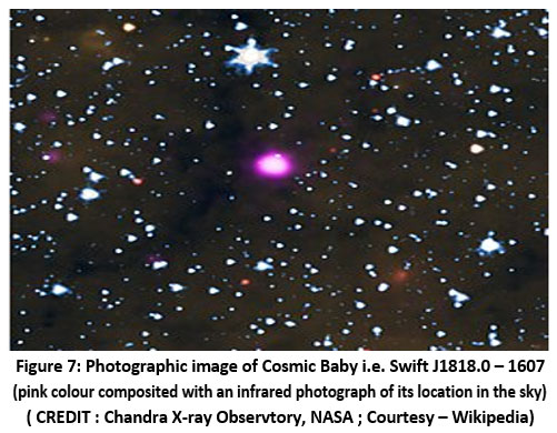 | Figure 7: Photographic image of Cosmic Baby i.e. Swift J1818.0 – 1607 (pink colour composited with an infrared photograph of its location in the sky ( CREDIT : Chandra X-ray Observtory, NASA ; Courtesy – Wikipedia) |
Another significant finding arises from the discovery of NANO Grav 35 – 37 which suggests that characteristics observed in cosmic objects , the same characteristics can also be possible to observe in human . Based on this Parui 38 suggested that triaxiality and other characteristics possibly be observable in human.
Crystallization of Protein in Magnetic field
Proteins play a significant role in governing almost all biochemical reactions in living things, particularly in human eye-lens from its structure formation to development. X-ray diffraction analysis shows that a protein single crystal is :
with length > 0.1 mm ;
of high quality is indispensible ;
with main problem is the crystallization that has been a bottleneck.
A possible solution to overcome these difficulties is investigation of the effect of magnetic field on protein crystallization. In an investigation of hen-egg white lysozyme as model protein and its effect on magnetic field 39 it was found that magnetic field effects on protein crystal is really a complex which needs a large quantity of high quality single protein crystal in order to understand the realistic characteristics of protein behavior, particularly on the orientation, crystal habit, growth rate of crystal, convection in aqueous solution, etc. In other words, effects of magnetic field and its magnetization force 40,41 on protein crystal offers an opportunity to the scientists a possible clues of high-resolution structural information that ultimately leads to high quality protein crystals.
Discussion
Protein crystallization in living cells 42, 43, particularly in human eye-lens plays a crucial role not only for beautiful eyes but also its various side-effects that create constraints via eye-diseases for clear vision. Investigations 44 – 46 of magnetic field effects on protein crystal growth provide possible clues to the scientists for obtaining high quality protein crystals that is required for clear vision’s tenure for a long years. Discovery of Cosmic Baby hints that deformation due strong magnetic field in the form of “Triaxiality” is applicable for human eyes also . This implies that at the early birth phase of zygote formation the human embryo may turn it’s shape into “Triaxility” when the gestational sac follow a shape change from circular to ellipsoidal. Thus, “Triaxiality” arises during the transition from circular to ellipsoidal shape of the gestational sac (that covers the embryo inside the mother’s womb) provides an invaluable opportunity to the scientists for investigating the exact role of “Triaxility” for ensuring formation of beautiful eyes as well as ageless appearance in women’s body structure.
Acknowledgement
The author is greatly indebted to the anonymous reviewers for their invaluable comments and suggestions. The author wishes to thank Prof. H N K Sarma, Dept. of Physics, Manipur University, B K Ganguly, Mrs. Tapati Parui and specially to Rajarshi Parui for his help in computer works.
Funding Sources
The author(s) received no financial support for the research, authorship, and/or publication of this article
Conflict of Interest
The author does not have any conflict of interest.
Data Availability Statement
Data sharing is not applicable as no data sets were analyzed.
Ethics statement
This research did not involve human participants, animal subjects, or any material that requires ethical approval.
Informed
This study did not involve human.
Author contributions
I have done the complete manuscript.
References
- Andley U P. Crystallins in the eye: Function and Pathology. Prog. Retinal & Eye Res. 2007 ; 26(1) : 78-98);
CrossRef - Bhat S P, Nagineni C N. Alpha B subunit of lens-specific Protein alpha crystallin is present om other ocular and non-ocular tissues. Biochem. Biophys. Res. Commun. 1989 ; 158 : 329 -338
CrossRef - Chayen N.E., Helliwell J.R., Snell E.H. : Macromolecular Crystallization and Crystal Perfection ( Oxford University Press : Oxford, UK . 2010).
CrossRef - Park S H, Suh S W , Song H. A cytosine modification mechanism revealed by the structure of a ternary complex of deoxycytidylate hydroxymethylase from bacteriophage T4 with its cofactor and substrate. IUCRJ 2019; 6 (2) : 206 - 217
CrossRef - Hong B, Haddad M, Maley F, Jensen J H, Kohen A. Hydride transfer versus hydrogen radical transfer in thymidylate synthase. J. Am. Chem Soc. 2006 ; 128 (17) : 5636 -5637
CrossRef - Cai R, Yang H, He J, Zhu W. The effects of magnetic fields on water molecular hydrogen bonds. J. Mol. Struct. 2009 ; 938 (1-3) : 15 - 19
CrossRef - Chang K.-T., Weng C-I. The effect of an external magnetic field on the structure of liquid water using molecular dynamics simulation. J. Appl. Phys. 2006 ; 100 : 043917
CrossRef - Adams PD, Afonine PV, Baskaran K, Berman H M, Berrisford J, Bricogne G, Brown D G, Burley S K, Chen M, Feng Z, Flensburg C, Gutmanas A, Hoch J C, Ikegawa J, Kengaku Y, Krissinel E, Kurisu G, Liang Y, Liebschner D, Mak L , Markley J L, Moriarty N W, Murshudov G N, Noble M, Peisach E, Persikova I, Poon B K, Sobolev O V, Ulrich E L, Velankar S, Vonrhein C, Westbrook J, Wojdyr M, Yokochi I M, Young J Y. Announcing mandatory submission of pdbx/mmcif format files for crystallographicdepositions to the protein data bank (PDB). Acta Cryst. D Struct Biol. 2019 ; 75 : 451 -545
CrossRef - Burley S K, Berman H M , Kleywegt G J, Markley J L , Nakamura H, Velankar S. Protein data bank (PDB) :The single global macromolecular structure archive. Methods Mol. Biol. 2017 ; 1607 : 627 - 641
CrossRef - Takeda M, Miyanoiri Y, Terauchi T, Yang C J, Kainosho M. Use of h/d isotope effects to gather information about hydrogen bonding and hydrogen exchange rates. J. Magn. Reson. 2014 ; 241 (4) : 148-154
CrossRef - Hodge E A, Benhaim M A, Lee K K. Bridging protein structure, dynamics, and function using hydrogen/deuterium-exchange mass spectrometry. Protein Sci. 2020 ; 29 (4) : 843-855
CrossRef - Luft J R, Wolfley J R, Said M I , Nagel R M, Lauricella A M , Smith J L , Thayer M H , Veatch C K , Snell E H , Malkowski M G , Detitta G T . Efficient optimization of crystallization conditions by manipulation of drop volume ratio and temperature. Protein Sci. 2007 ; 16 (4) : 715 - 722
CrossRef - Asai T, Suzuki Y, Sazaki G, Tamura K, Sawada, T, Nakajima, K. Effects of high pressure on the solubility and growth kinetics of monoclinic lysozyme crystals. Cell. Mol. Biol. 2004 ; 50 : 329 -334
CrossRef - Hammadi Z, Veesler S. New approaches on crystallization under electric fields. Prog. Biophys Mol. Biol. 2009 ; 101(1-3) : 38 - 44
CrossRef - Dong J, Boggon T J, Chayen N E, Raftery J, Bi R C, Helliwell J R. Bound-solvent structures for microgravity-, ground control-, gel- and microbatch-grown hen egg-white lysozyme crystals at 1.8 a resolution. Acta Cryst. D Biol. Cryst. 1999 ; 55 (Pt-4) : 745 - 752
CrossRef - Pareja-Rivera C, Cuéllar-Cruz M, Esturau-Escofet N, Demitri N, Polentarutti M , Stojanoff V, Moreno A. Recent advances in the understanding of the influence of electric and magnetic fields on protein crystal growth. Cryst. Growth Des. 2016 ; 17 (1) : 135- 145
- Hünefeld F L. Der Chemismus in der thierischen Organisation , Leipzig: Brockhaus, 1840 ; p. 158-63
- Hartig T. Ueber das Klebermehl. Botanische Zeitung 1855 ; 13 : 881 - 882
- Osborne T B. Crystallized vegetable proteids. Amer. Chem. J. 1892 ; 14 : 662 - 690
CrossRef - Parui R K. Cosmic Baby and Crystallography — Decoding the Invisible inside the Secret of the Universe. Orient. J. Phys. Sci. 2024 ; 09 ,
- Reichert E T, Brown A P. The Differentiation and Specifity of Corresponding Proteins and other vital substances in relation to biological classification and evolution, in The Crystallization of Hemoglobins. (Carnegie Institution, Washington, DC. 1909)
CrossRef - Ehrnsperger M, Graber S, Gaestel M, Buchner J. Binding of non-native protein to Hsp25 during heat shock creates a reservoir of folding intermediates for reactivation. EMBO J. 1997 ; 16 (2) : 221 - 229
CrossRef - .Jakob U, Gaestel M, Engel K, Buchner J. Small heat shock proteins are molecular chaperones. J Biol Chem. 1993 ; 268 (3) : 1517 - 1520
CrossRef - Lee G J., Roseman A.M, Saibil H.R., Vierling, E. A small heat shock protein stably binds heat-denatured model substrates and can maintain a substrate in a folding-competent state. EMBO J. 1997 ; 16 : 659 - 671
CrossRef - Peschek J, Braun N, Franzmann T M, Buchner J. The eye lens chaperon ?-crystallin forms defined globular assemblies. PNAS. 2009 ; 106 (32) : 13272 - 13277
CrossRef - Preston G M , Carroll T P, Guggino W B, Agre P. Appearance of water channels in Xenopus oocytes expressing red cell CH1P28 Protein. Science 1992 ; 256 : 385
CrossRef - Agre P. Aquaporin Water Channels , Nobel Lecture 8 December 2003 (Johns Hopkins University School of Medicine, Bultimore, MD. ) p. 184
- Gehrels N , Chincarini G, Giommi P, Mason K O, Nousek JA, Wells AA, White N E , Barthelmy S D, Burrows D N, Cominsky L R, Hurley, K C, Marshall FE, Mészáros P, Roming P W A, Angelini L., Barbier L M, Belloni T, Campana S, Caraveo PA., Chester M M, Citterio O, Cline T L, Cropper M S, Cummings J R, Dean A J, Feigelson E D, Fenimore E E, Frail D A, Fruchter A S, Garmire G P., Gendreau K, Ghisellini G, Greiner J, Hill J E, Hunsberger S D, Krimm H A, Kulkarni S R, Kumar P, Lebrun F, Lloyd-Ronning N.M., Markwardt C B, Mattson B J, Mushotzky R F, Norris J P, Osborne J, Paczynski B, Palmer D M, Park H.-S, Parsons A M, Paul J, Rees J M, Reynolds C S, Rhoads J E, Sasseen T P, Schaefer B E, Short A T, Smale A P, Smith L A, Stella L , Tagliaferri G, Takahashi T, Tashiro M, Townsley L K, Tueller J, Turner M J L, Vietri M, Voges W, Ward M, Willingale R, Zerbi F M, Zhang W W. The Swift Gamma Ray Bursts Mission. Astrophys. J. 2004 ; 611 : 1005
CrossRef - Gehrels N, Razzaque S. Gamma Ray Bursts in the Swift Fermi Era. Frontiers Phys. 2013 ; 8 : 661
CrossRef - Evans P A, Gropp J D, Kennea J A, Klingler N J, Laha S, Lien A Y, Page K L, Sakamoto T, Tohuvavohu, A, Neil Gehrels Swift Observatory Team. Swift BAT trigger 960986 : Swift detection of a new SGR Swift J 1818.0 – 1607. 2020 ; GCN Circular # 272273
- Esposito P, Rea N, Borghese A, Coti Zelati F, Viganò D, Israel G L , Tiengo A, Ridolfi A, Possenti A , Burgay M, Götz D, Pintore F, Stella L, Dehman C, Ronchi M, Campana S, Garcia-Garcia A, Graber V, Mereghetti S, Perna R, Rodríguez Castillo G A, Turolla R, Zane S. A very young radio-loud Magnetar. Astrophys. J. Lett. 2020 ; 896 : L30
CrossRef - Parui R K. A Remark on “Do triaxial star supermassive compact star exist ? Int. Astron. Astrophys. Res. J. 2023 ; 5 (1) : 33-37
- Parui R K. A New Compact Star – the “Triaxial Star” – and the Detection of a Cosmic Baby: A Possibility. Int. Astron. Astrophys. Res. J. 2023 ; 5 (1) : 38 - 47
- Parui R K. Cosmic Baby and detection of a new compact Star – the “Triaxial Star” : A possibility. Astrophys. Space Sci. 2023 ; 368 (6) : 46 . Revised to be submitted (2025)
- Chandrasekhar S. Ellipsoidal Figures of Equilibrium. (Yale Univ. Press. New Haven, USA ,1969)
- Nano Grav Team : Agazie G, Anumarlapudi A, Archibald A M , Baker P T, Becsy B, Blecha L, Bonilla A, Brazier A, Brook P R, Burke-Spolaor S, Burnette B , Case R, Casey-Clyde J A, Charisi M, Chatterjee S, Chatziioannou K, Cheeseboro B D, Chen S, Cohen T, Cordes J M, Cornish N J , Crawford F, Cromartie H T, Crowter K, Cutler C J , D'Orazio D J, DeCesar M E, DeGan D, Demorest P B, Deng H, Dolch T, Drachler B, Ferrara E C, Fiore W , Fonseca E , Freedman G E , Gardiner E, Garver-Daniels N, Gentile P A, Gersbach KA, Glaser J, Good D C, Gültekin K, Hazboun J S, Hourihane S, Islo K, Jennings R J, Johnson A, Jones M L, Kaiser A R , Kaplan D L , Kelley L Z, Kerr M, Key J S, Laal N, Lam M T , Lamb W G, Joseph T, Lazio W, Lewandowska N , Littenberg T B , Liu T, Luo J, Lynch R S, Ma C-P, Madison D R, McEwen A, McKee M W, McLaughlin M A, McMann N, Meyers B W , Meyers P M , Mingarelli C M, Mitridate A, Natarajan P, Ng C, Nice D J, Ocker S K , Olum K D, Pennucci TT , Perera B B P, Petrov P, Pol N S , Radovan H A , Ransom S M , Ray P S, Romano J D , Runnoe J C, Sardesai S C, Schmiedekamp A, Schmiedekamp C, Schmitz K, Schult L, Shapiro-Albert B J , Siemens X, Simon J, Siwek M S, Stairs I H, Stinebring D R, Stovall K, Sun J P, Susobhanan A, Swiggum J K, Taylor J, Taylor S R, Turner J E, Unal C, Vallisneri M, Vigeland S J, Wachter J M , Wahl H M, Wang Q, Witt C A, Wright D, Young O, and The NANOGrav Collaboration.. The NANOGrav 15 yr Data set : Constraints on Supermassive Black hole Binaries from the gravitational wave background. Astrophys. J. Lett. 2023 ; 952 : L37
CrossRef - Nano Grav. Team. Agazie G, Anumarlapudi A, Archibald A M, Baker P T, B. Becsy, B. Blecha, Blecha L, Brazier A, Brook P R, Burke-Spolaor S, Burnette R, Case R, Charisi M, Chatterjee S, Chatziioannou K, Cheeseboro B D, Chen S, Cohen T, Cordes J M, Cornish N J , Crawford F, Cromartie H T , Crowter K, Cutler C J, DeCesar M E, DeGan D, Demorest P B, Deng H, Dolch T, Drachler B, Ellis J A, Ferrara E C , Fiore W, Fonseca E, Freedman G E, Garver-Daniels N, Gentile P A,Gersbach K A, Glaser J, Good D C, Gultekin K, Hazboun J S, Hourihane S, Islo K, Jennings R J, Johnson, A D, Jones M L, Kaiser A R, Kaplan D L, Kelley L Z, Kerr M, Key J S, Klein T C, Laal C, Lam M T, Lamb W G, Joseph T, Lazio W, Lewandowska N, Littenberg T B, Liu T, Lommen A, Lorimer D R, Luo J, Lynch R S, Ma C-P, Madison D R, Mattson M A, McEwen A , McKee J A, McLaughlin M A, McMann N, Meyers B W, Meyers, P M, Mingarelli C, Mitridate A, Natarajan, Ng C, Nice D J, Ocker S K, Olum K D , Pennucci T T , Perera B B P, Petrov P, Pol N S, Radovan H A, Ransom S M, Ray P S , Romano J D , Sardesai S C, Schmiedekamp A, Schmiedekamp C, Schmitz K, Schult L, Shapiro-Albert B J , Siemens X, Simon J, Siwek M S , Stairs I H , Stinebring D R , Stovall K , Sun J P, Susobhanan A, Swiggum J K , Taylor J, Taylor S R , Turner J E, Unal C, Vallisneri M, van Haasteren R, Vigeland S J, Wahl H M , Wang Q, Witt C A , Young O. The NANOGrav 15 yr Data Set: Evidence for a Gravitational-wave Background . Astrophys. J. Lett. 2023 ; 951: L8
CrossRef - Nano Grav Team : Agazie G, Alam M F , Anumarlapudi A, Archibald AM , Arzoumanian Z, Baker P T, Blecha L , Bonidie V, Brazier A, Brook P R , Burke-Spolaor S, Bécsy B, Chapman C, Charisi M, Chatterjee S, Cohen T, Cordes J M, Cornish N J , Crawford F , Cromartie H T, Crowter K , DeCesar M E , Demorest P B , Dolch T , Drachler B, Ferrara E C, Fiore W, Fonseca E , Freedman G E , Garver-Daniels N, Gentile P A , Glaser J, Good D C , Gültekin K , Hazboun J S , Jennings R J, Jessup C, Johnson A D, Jones M L , Kaiser A R , Kaplan D L , Kelley K Z , Kerr M, Key J S , Kuske A, Laal N, Lam M T , Lamb W J , Lazio T J W, Lewandowska N, Lin Y, Liu T, Lorimer D R , Luo J, Lynch R S, Ma C P , Madison D R , Maraccini K , McEwen A, McKee J W , McLaughlin M A , McMann N, Meyers B W, Mingarelli C M F, Mitridate A, Ng C, Nice D J , Ocker S K , Olum K D , Panciu E , Pennucci T T , Perera B B P , Pol N S , Radovan H A , Ransom S M , Ray P S , Romano J D , Salo L, . Sardesai S C , Schmiedekamp C, Schmiedekamp A, Schmitz K, Shapiro-Albert B J , Siemens X, Simon J, Siwek M S , Stairs I H , Stinebring D R, Stovall K , Susobhanan A, Swiggum J K , Taylor S R , Turner J E , Unal C, Vallisneri M, Vigeland J, Wahl H M, Wang Q, Witt C A , Young O, and The NANOGrav Collaboration. The NANOGrav 15 yr Data Set: Observations and Timing of 68 Millisecond Pulsars. Astrophys. J. Lett. 2023 ; 951 : L9
CrossRef - Parui R K. Cosmic Baby and Human Baby — Do both of them exhibit a similar type characteristics as cosmic connection to human ? : A possibility . Revised version submitted to Euro. Phys. J. C (2024)
- Sazaki G, Yanagiya S, Durbin S D, Miyashita S , Nakada T, Komatsu H, Ujihara T, Nakajima K, Watanabe K , Motokawa M. Effects of a Magnetic field on the Crystallization of Protein. in Materials Science in Static High Magnetic fields, eds: M Motokawa , K. Watanabe . Springer-Verlag, Berlin Heidelberg. 2002 , p. 283
CrossRef - Alka M, Wakayama N I. Effects of a magnetic field and magnetization force on Protein crystal growth: Why does a magnet improve the quality of some crystals ? Acta Cryst. D 2002 ; 58 : 1708 - 1710
CrossRef - Alka M. The influence of magnetic field on protein crystal growth and quality. Deutsches Elektronen Synchrotron (DESY), Hamburg, Germany (2005)
- Schonherr R, Rudolph J M, Redecke L. Protein crystallization in Living Cells. Biol. Chem. 2018 ; 339 (7) : 751- 772
CrossRef - Jiang M, Zhang L, Wang F, Zhang J, Liu G, Gao B, Wei D. Novel application of Magnetic protein: Convenient one-step purification and immobilization of proteins. Nature (Scientific Reports) 2017 ; 7 (1) : 13329 -13329
CrossRef - Ryu S Y, Oh I H, Cho S J, Shin A K, Hyun K S. Enhancing protein crystallization under a Magnetif field, Crystals 2020 ; 10 (9) : 821
CrossRef - Schey K L, Wang Z, Wenke J L, Ai Y . Aquaporins in the eye: Expression, function, and roles in Ocular disease. Biochim. Biophys. Acta. 2014 ; 1840 : 1513 - 1523
CrossRef - Surade S, Ochi T, Nietlispach D, Chirgadze D , Moreno A. Investigation into Protein Crystallization in the presence of a strong magnetic field. Crystal. Growth. Design. 2010 ; 10 : 691- 699
CrossRef

This work is licensed under a Creative Commons Attribution 4.0 International License.